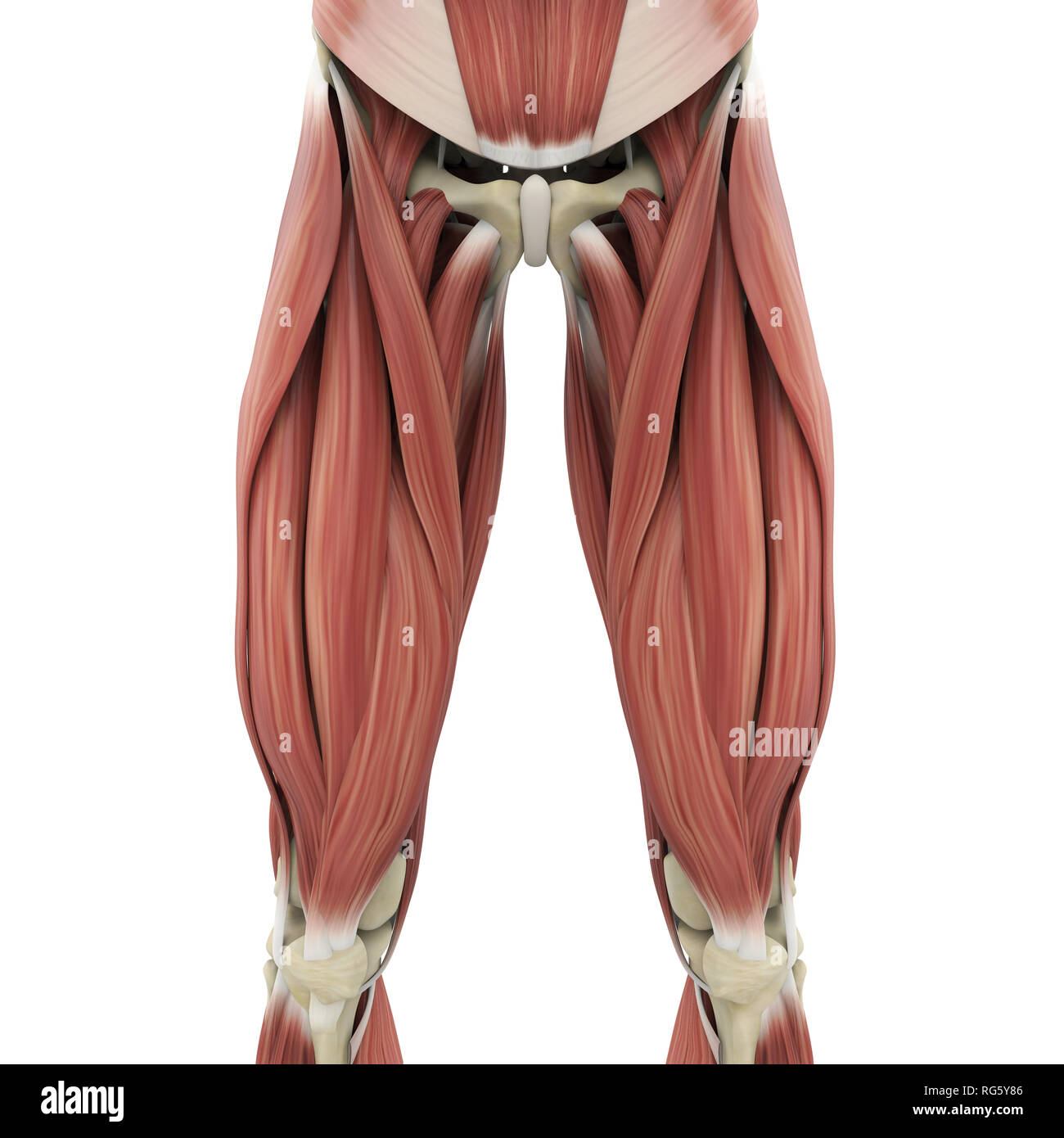
Upper Thigh Anatomy - Muscles of the anterior thigh. Muscles of the arm and forearm diagram. Bf sh, lh, biceps femoris short head, long head; These images are a random sampling from a bing search on the term thigh anatomy. click on the image (or right click) to open the source website in a new browser window. We are pleased to provide you with the picture named upper thigh muscle anatomy.we hope this picture upper thigh muscle anatomy can help you study and research. Muscle adductor thigh anatomy fiber pectineus psoas upper body human longus tendon 3d athlete biology bodybuilding bone femoris fitness foot gracilis health iliacus iliotibial illustration ilopsoas isolated knee lateralis leg ligament male man medical medicine model muscular patella quadriceps rectus.
They originate at the ilium (upper part of the pelvis, or hipbone) and femur (thighbone), come together in a tendon. Clinical anatomy for dummies cheat sheet. Pelvic & upper thigh anatomy. In human anatomy, the thigh is the area between the hip (pelvis) and the knee. We look at the associated symptoms and treatment options.
Muscles of the Thigh Part 1 - Anterior Compartment Anatomy - YouTube from i.ytimg.com Vascular anatomy of the upper arm. In human anatomy, the thigh is the area between the hip (pelvis) and the knee. Want to learn more about it? Upper part of the ischial tuberosity insertion: The information contained in anatomy atlases is not a substitute for the medical care and advice of your physician. Bf sh, lh, biceps femoris short head, long head; We are pleased to provide you with the picture named upper thigh muscle anatomy.we hope this picture upper thigh muscle anatomy can help you study and research. Its bones, muscles, nerves, joints, blood vessels and lymphatics, anatomical areas, and structures found.
Bf sh, lh, biceps femoris short head, long head;
Tibial part of the sciatic nerve action: Anatomy of the human body. This webpage presents the anatomical structures found on thigh mri. The single bone in the thigh region is called the femur. The axilla and the deltoid region in axial and coronal and axial. Other articles where thigh is discussed: These images are arranged in radiographic view, as though you were looking up from the patient's feet toward the head. The anatomical areas found on the upper limb can serve as key landmarks to help us find important anatomical structures such as finding one of the in this section, learn more about the upper limb: Forearm anatomy upper limb anatomy anatomy study anatomy reference pose reference gross anatomy human body anatomy human anatomy thigh muscle anatomy leg muscles anatomy muscular system anatomy hip anatomy human muscle anatomy human anatomy drawing. It passes obliquely across the upper and anterior part of the thigh, from the lateral to the medial side of the limb, then descends vertically, as far as the medial side of the knee, passing behind the medial condyle of the. The information contained in anatomy atlases is not a substitute for the medical care and advice of your physician. Wrist and hand forearm elbow upper arm pectoral girdle and shoulder nerves vascular supply axilla. Its bones, muscles, nerves, joints, blood vessels and lymphatics, anatomical areas, and structures found.
Forearm anatomy upper limb anatomy anatomy study anatomy reference pose reference gross anatomy human body anatomy human anatomy thigh muscle anatomy leg muscles anatomy muscular system anatomy hip anatomy human muscle anatomy human anatomy drawing. The single bone in the thigh region is called the femur. …front and sides of the thigh. Anatomy atlases, the anatomy atlases logo, and a digital library of anatomy information are all trademarks of michael p. We are pleased to provide you with the picture named upper thigh muscle anatomy.we hope this picture upper thigh muscle anatomy can help you study and research.
Elbow/Radioulnar Joint & Knee Joint Muscles & Actions | Tiger Training from tigerfitandwell.files.wordpress.com There may be variations in treatment that. This is a great exercise for toning your upper thigh muscles. Anatomically, it is part of the lower limb. In human anatomy, the thigh is the area between the hip (pelvis) and the knee. Other articles where thigh is discussed: Related posts of muscle anatomy of upper thigh. It passes obliquely across the upper and anterior part of the thigh, from the lateral to the medial side of the limb, then descends vertically, as far as the medial side of the knee, passing behind the medial condyle of the. Muscle adductor thigh anatomy fiber pectineus psoas upper body human longus tendon 3d athlete biology bodybuilding bone femoris fitness foot gracilis health iliacus iliotibial illustration ilopsoas isolated knee lateralis leg ligament male man medical medicine model muscular patella quadriceps rectus.
These images were created using data obtained from the final chapter presents anatomical charts of anatomical sections of the upper limb:
Start studying thigh/upper leg anatomy. Bf sh, lh, biceps femoris short head, long head; Each arm consists of four main parts These images are arranged in radiographic view, as though you were looking up from the patient's feet toward the head. These images were created using data obtained from the final chapter presents anatomical charts of anatomical sections of the upper limb: Rectus femoris, vastus lateralis, vastus medialis, and vastus intermedius. Muscle anatomy 12 photos of the muscle anatomy human muscle anatomy video, muscle anatomy abdomen, muscle anatomy diagram, muscle anatomy rotator cuff, muscle anatomy website, human. Pelvic & upper thigh anatomy. Tibial part of the sciatic nerve action: This section of the website will explain large and minute details of arterial anatomy of upper legs (thigh arteries). …front and sides of the thigh. Anatomically, it is part of the lower limb. Anatomically, it is part of the lower limb.
Thus, the right side of the image is the patient's left. Clinical anatomy for dummies cheat sheet. Some clinical anatomy highlights of the thorax, abdomen, and pelvis. In human anatomy, the thigh is the area between the hip (pelvis) and the knee. These images are a random sampling from a bing search on the term thigh anatomy. click on the image (or right click) to open the source website in a new browser window.
Human Upper Leg Muscles High Resolution Stock Photography and Images - Alamy from c8.alamy.com Bf sh, lh, biceps femoris short head, long head; The muscles and fasciæ of the thigh. Illustrations of the anatomy of the upper limb. These images are a random sampling from a bing search on the term thigh anatomy. click on the image (or right click) to open the source website in a new browser window. Forearm anatomy upper limb anatomy anatomy study anatomy reference pose reference gross anatomy human body anatomy human anatomy thigh muscle anatomy leg muscles anatomy muscular system anatomy hip anatomy human muscle anatomy human anatomy drawing. Medial condyle of tibia nerve supply: Anatomically, it is part of the lower limb. The single bone in the thigh region is called the femur.
Clinical anatomy for dummies cheat sheet.
Each arm consists of four main parts …front and sides of the thigh. In human anatomy, the thigh is the area between the hip (pelvis) and the knee. Coronal arterial anatomy of upper legs (thigh). Anatomy atlases, the anatomy atlases logo, and a digital library of anatomy information are all trademarks of michael p. We look at the associated symptoms and treatment options. This webpage presents the anatomical structures found on thigh mri. They originate at the ilium (upper part of the pelvis, or hipbone) and femur (thighbone), come together in a tendon. Muscles attachment , anatomy atlas. As an artist, fitness instructor, master of nutrition student, and former massage therapist, i had to have totally unique, funky, and fresh anatomy charts for my study. • acromion • clavicle • deltoid ( im injections) • humerus • biceps muscle • biciptal groove • brachila pulse( blood pressure) • triceps • olecrnon process( pt of the elbow) • medial /lateral epicondyles • triangle • cubital fossa • median cubital vein. Medial condyle of tibia nerve supply: This section of the website will explain large and minute details of arterial anatomy of upper legs (thigh arteries).
Share :
Post a Comment
for "Upper Thigh Anatomy - Muscles of the anterior thigh"


Post a Comment for "Upper Thigh Anatomy - Muscles of the anterior thigh"